MRI
Interpreting MRI as environments/systems/interfaces
where body-machine and analogue-digital meet
︎︎︎
ABSTRACT
Corporeal matter as perceived by MRI straddles definitions of substance, organism, subject and object. MRI interacts with the body through nuclear magnetic resonance and electrodynamics, bringing us into contact with the body as a person, as an assemblage of biochemical reactions, as a patient and a cellular, molecular, atomic and subatomically composed entity.
This research project and exhibition use artistic processes as a method for investigating MRI and what it means to be a body as defined by MRI. The artworks exhibited were created as part of a process of interacting with the physics of MRI and making art objects as scientific devices.
Being largely isolated from how computational technologies work, there is an increasing incentive to make their internal processes visible. Particularly in a broader context where so much of the gig economy is exploitative and obscures the labour that produces electronics and services alike. The environmental, human and inter-species cost is also concealed behind the apparent seamlessness of big tech, AI and user experience design (see Anatomy of an AI). The lives lost in mines, extraction processes, factories and distribution hubs needs to be a central concern. How we communicate and exchange our ideas and feelings is also being shaped by these technologies, this is also a form of unpaid labour.
This exhibition begins with MRI and MRI data. The sculptural, woven, drawn and painted work exhibited draws on the mathematical and physical processes of MRI. The artwork exhibited makes use of scientific information and biomedical imaging laboratories as part of the creative practice.
The artwork exhibited includes sculptural ‘phantoms’ that are used to investigate the interface between body and machine in MRI and are named after scientific devices of the same name. By thinking about how matter interacts with MRI, I could make these artefacts from within and as part of MRI. My artistic phantoms are made using ‘tissue mimicking materials’ that from the perspective of the scanner, could be part of a living organism.
You will also find woven works, patterns and swatches created in response to the way in which an MRI scanner turns signals from the body into a digital image of the body. These woven works and the patterns developed for them are based on the mathematical processes of signal analysis. They were made from within and part of the MRI process. Drawings, illustrations, diagrams and paintings created as part of my research process are also created to chart my artistic practice's entanglements and constellational nature.
PROJECT
OVERVIEW
This website presents artworks I created as part of my practice-based PhD project carried out at the University of Portsmouth. This research project began with MRI and makes use of scientific knowledge and personal experiences of medicine and health care.
Being largely isolated from how computational technologies work, there is an increasing incentive to make their internal processes accessible.
Through this research project and the experience of making the artefacts exhibited, I developed embodied approaches to understanding biomedical imaging that explore the ways in which MRI data is organised and codified. My sculptural, woven, drawn and painted practices draw on the mathematical and physical processes of MRI.
Sculptural ‘phantoms’ investigate the interface between body and machine and are semi-figurative constructs informed by how the body interacts with the physics of MRI. I also developed a weaving practice as an embodied method to investigate how analogue signals in MRI are transformed into digital biomedical images. My woven work is a non-pictorial deconstructive reconfiguration of mathematical phenomena needed in signal analysis. My aim was to investigate through artistic creative practice how this aspect of MRI technology works.
The creative acts of drawing, illustration, making diagrams and paintings chart the entanglements of the body, bodily materials, and their interactions with MRI. The visuals and images produced also act as a map connecting the various elements of my art practice to my findings, materials, processes, places, systems, environments and my experience of being a cancer patient.
THE MRI PROCESS
MRI is a non-invasive biomedical imaging technology that visualises tissues within the body. MRI is an interesting piece of physics that interacts with the body. As a technology it draws on the quantume mechanical properties embedded in corporeal matter - particularly hydrogen ions, also known as protons (H+). MRI brings together multiple aspects of our ontology. MRI interacts with the body through nuclear magnetic resonance and classical electrodynamics and thus physics allows us to connect to the abject. This brings us into contact with the body as a person, a patient, a member of a community, and as a cellular, molecular, atomic and subatomically composed entity.
The magnetic field of an MRI penetrates our materiality and requires both quantum mechanics and classical electrodynamics to work. MRI involves the phenomena of nuclear magnetic resonance (NMR) and the intrinsic subatomic properties of spin, angular momentum and magnetism possessed by atomic nuclei.
Protons within atomic nuclei are like tiny spinning magnets. The first stage of MRI involves the powerful MRI magnet which aligns the spin of the protons, lining them up like synchronised swimmers. Following this alignment, the protons are energised with radio frequency electromagnetic energy (RF photons) in the form of high-frequency RF pulses. This causes them to resonate.
As the protons absorb the RF energy, they wobble away from the magnet’s alignment to varying degrees depending on what material or substance they are in. When the RF signal is turned off the protons still resonate. Refocusing the magnet re-aligns the protons, causing them to re-emit the difference in RF as the NMR signal.
The time it takes for the protons to emit the signal is called relaxation time; it generally comes in two kinds of measurement: T1 and T2. The difference in relaxation time and proton density are essential factors in creating accurate MRI images. When the NMR signal is emitted it is detected in the scanner. These signals are converted into a digital biomedical image using the mathematics of signal analysis including a type of maths called Fourier transforms (FTs). This process is computational and mathematical where the wave properties of the NMR signal are used to figure out what kinds of matter/molecules are resonating where.
The body and its substances, molecules and atoms interact with the MRI system (magnet, RF signals and sensors/detection mechanisms) at the ‘body machine interface’. This interface is beyond (or within) the boundary of the skin. The magnet and RF signals are able to work across multiple scales of magnitude. By thinking about MRI in this way I was able to create sculptures that interact with MRI as if it were a body. Making sculptures with the body-machine interface in mind made it possible for my work to intersect, interact and intra-act with MRI. The analogue NMR signal being converted/processed/computationally interpreted into a digital biomedical image is perceived as the analogue-digital interface in my research (a non-scientific term).
An interface is the means by which interaction or communication is achieved or is a surface forming a common boundary between two or more bodies, spaces, or phases. Interface is a verb and a noun that entails action: to connect or become interfaced. In my making processes I consider the behaviour of corporeal matter at these interfaces and the types of connection, disconnection and reconnection that take place. Understanding what happens at the interfaces helps me decide how to develop practices that interact and interface with MRI on the molecular and mathematical levels.
MRI
TRIALS
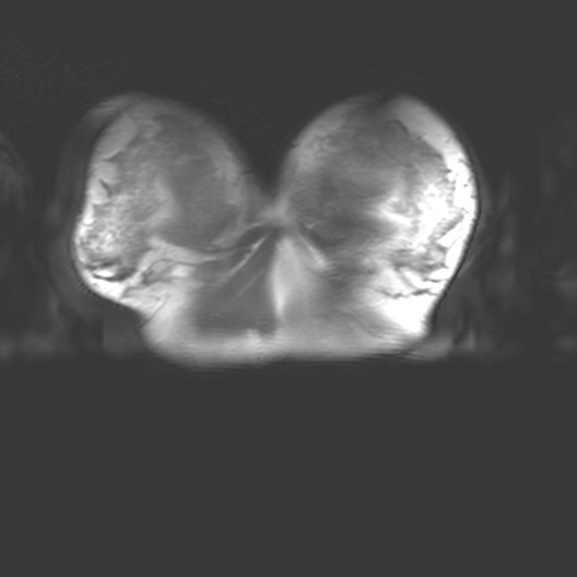
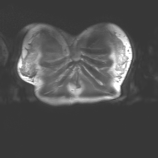
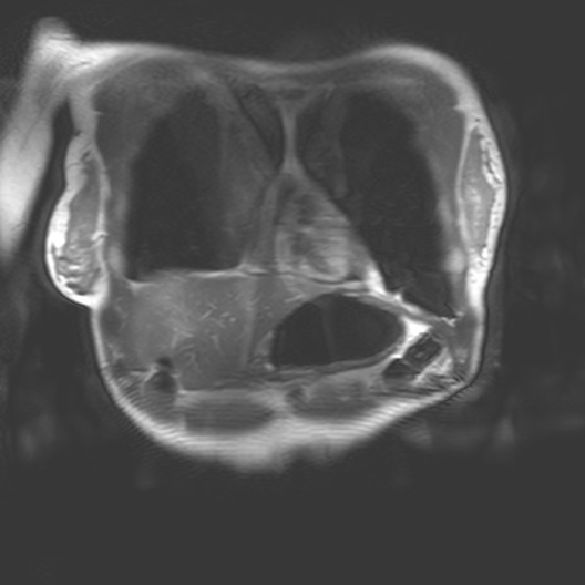
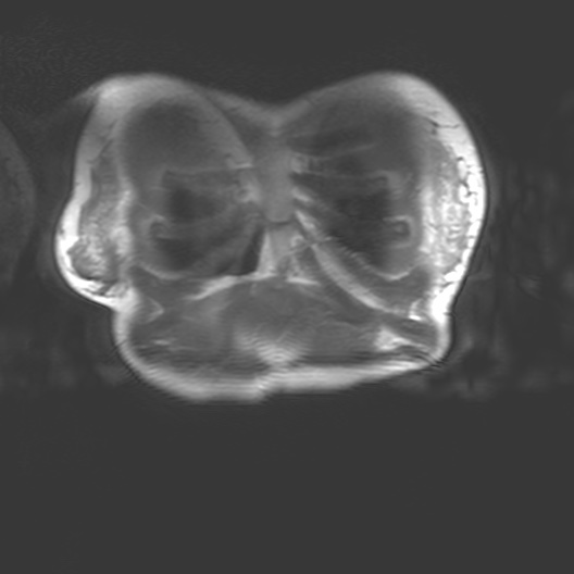
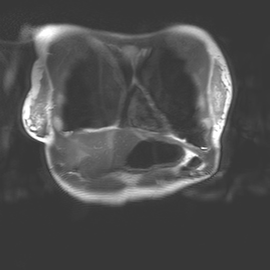
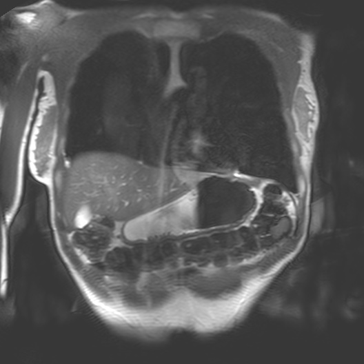
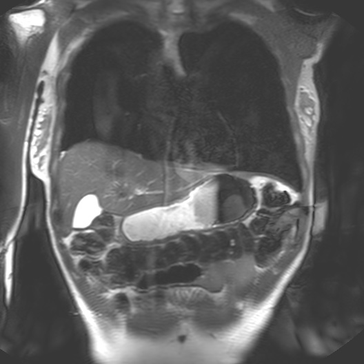
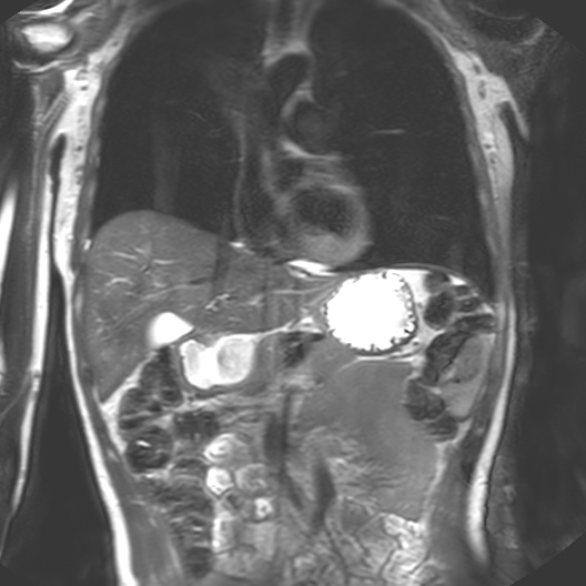
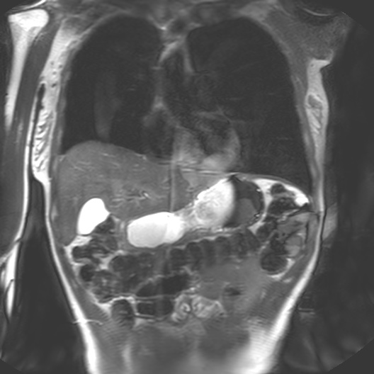
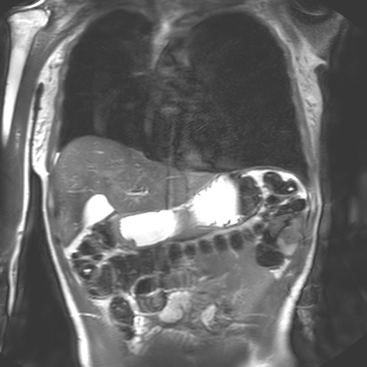
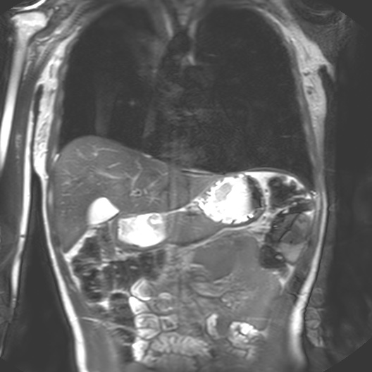
I began my investigation by taking part in MRI trials at the Cancer Centre in 2018 with Dr Heather Fitzke. MRI images confront us with the fact that our bodily materiality acts beyond us and that medical technology shapes our sense of self.
Seeing my organs autonomously pulsing on the monitor in the control room had a pronounced impact on me. It was like looking into a rockpool within my own body. The affecting power of MRI (its power to change our emotions) is in its potential to reveal autonomic bodily functions and diseases which are beyond our control. The data on the screen revealed my autonomous self: peristalsis, bowel movements, digestion, heartbeats and respiration. Through MRI, anatomy is seen as embedded and relational.
I felt a deep sense of fascination with how my organs moved and worked. Haraway’s situated biopolitics argues for an account of the body as an environment, in a state of simultaneous dissolution and formation which I notice in my data. My organs seemed like invertebrates in a rockpool: a squishy pulsing ecosystem. Organs do not operate in isolation but are interconnected. Their autonomous pulsing and motion seemed creaturely and strange in contrast to my numerous encounters with preserved cadavers in the dissecting room. My organs keep me alive yet I have no conscious sense of their functioning. They function beyond me.
Being the subject of MRI was an opportunity to experience how to interface or interact with it. One of my research aims concerns the materialisation of data processes in the MRI system that are not visible or accessible to the subject. The ‘hidden’ processes at the body-machine interface concern the interaction of corporeal matter with nuclear magnetic resonance phenomena (NMR).
Selecting the correct materials to make phantoms was the first step in interacting with the body-machine (interface). I would refer back to my data sculpture and cross-sectional projections from my scans with Dr Fitzke.
Anatomy as a rockpool
![]()
![]()
![]()
![]()
![]()
![]()
![]()
![]()
![]()
![]()
![]()
![]()
![]()
![]()
![]()
![]()
![]()
![]()
![]()
![]()
![]()
![]()
![]()
![]()